I am sure much of you think the image stops in the eye, that is, that this is where the process ends. We see with the eyes, don’t we? But maybe, after you read the previous post, your thoughts are confused.
In the other hand, I am sure some of you know that the brain is involved in this process but you do not know or understand very well how the connection is between eyes and brain.
It is true that this process is complicated ENOUGH, that is why, so it is easier to understand, I will show how what we see is processed from a practical example and with some graphics.
 Let’s suppose we are driving on a street and we are arriving to a cross. In the corner we see the “STOP” traffic sign.
I am going to show you how much information is processed just when seeing this simple traffic sign.
- In the first instance, we have the VISUAL PROCESS itself, that is, obtaining the clear image.
If you remember the previous post, the image created in both eye foveas is processed by the cones (A) that are in the central area of the retina. They send the information to the parvo ganglion cells (C) and leave the eye across the optic nerve (11) . These ganglion cells take the information about the color, bright, clarity and contrast of the STOP traffic sign to the LATERAL GENICULATE NUCLEUS. This is a relief and control station in the visual pathway: almost all information -that the parvo ganglion cells receive-arrives here, and this structure filters and leads it towards the respective places in the brain. So, only ten percent of this visual information is sent to the OCCIPITAL LOBE or VISUAL CORTEX, in order to create the image.
This particular process is incredibly much more complex, but by now, this is enough to understand the rest of the visual information processing
______________________________________
The rest of the pathways that take part in this visual information processing, are not called “visual pathways”, because the information does not go to the occipital lobe directly. The following information that is processed, is not purely visual, although the information had been obtained through the eyes.
- Therefore, in the second instance, we have the following PROCESS: EYES -> SUPERIOR COLLICULUS -> PARIETAL LOBE. In this process the information about “Where am I?” is obtained:
This pathway is made up basically of magno ganglion cells (C) that received the information from the rods in both retinas.
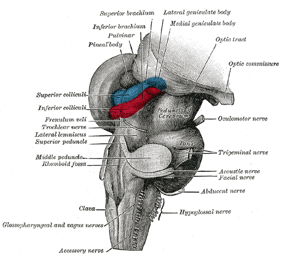
The Superior Colliculus is a paired structure of the nervous system placed in the mesencephalon (2) , right under the thalamus. The Superior Colliculus belongs to the Brainstem, which is the structure that joins the Brain and the Spinal Cord.
It deals to integrate the visual input with the auditory, somaticsensory (about balance and proprioception, among others) and tactile input; and thus we achieve the information about “Where are we?”.
In order to answer this question, the Superior Colliculus makes eyes and head move automatically towards the stimuli in the environment.
In the practical example, the group of all information is processed so that the Posterior Parietal Lobe allows us to calculate our movement, the speed of our car, the strength that we need to do in order to put our foot on the brake pedal, the direction of our car, or the rest of the cars, where our feet are, where our hands are, where we have to stop near the traffic sign… This lobe give us a metal spatial map about ourselves.
Therefore, when we have a car crash with a collision from behind, our head suffers a “lash” front to back, and in this case, the part of the brain is usually is affected is the mesencephalon, so, as well as we suffer strong cervical pains after the accident, it is also frequent to suffer disorientation, poor concentration, panicky feeling in places with crowd or difficulty for going up or down stairs. These are symptoms that seems light and that many people might not attribute to the car crash, but they make their daily life difficult. We can help these people by using Vision Therapy to get back the lost stability back.
- In the third instance, we have the following PROCESS: OCCIPITAL LOBE -> -> POSTERIOR PARIETAL LOBE. In this process the information about “Where is the object?” -the traffic sign-, is obtained:
The information from magno ganglion cells that arrives to the occipital lobe, does not stop here; some of these mango ganglion cells (C) go to Medial Temporal Lobe and, from there, go to the posterior parietal lobe to give us some information about “Where is the stop?”: Where is the line? Where must we stop? Where is the brake pedal to put our foot? Where is the car behind us or in front us?... As I explained above, Parietal Lobe allows us to make special calculations, as in the previous process. This lobe gives us a mental spatial map about our environment.
The information about “Where am I?” and “Where is the object?” allows the parietal lobe to make a motor plan, that is, “how we must do the things”. This lobe allows us to look at the road in the mean time: we look at the mirrors, we turn the wheel if it is necessary, we keep on the security distance regarding the front car, turn on the headlights if we get into a tunnel, we talk to another person in the car, we listen to the radio,...
But if you think about it, this process is carried out unconsciously, that is, all information that comes from the peripheral retina (Magno system) is processed and is carried out automatically. The same thing happens when we are driving on a road and we find an obstacle in our lane; then we look at the rear mirror and if there is not any further risk, we slightly turn the wheel to avoid it.
The responsible lobe for carrying out this action is the parietal one, but we have to do that unconsciously, as a reflex.
When we want to pass another car in the road and we have another one in the opposite direction, this lobe gives us some information about the following: what speed is the other car going at? What speed is the front car going at? What speed is our car going at? What car is moving faster? Do we have enough time to pass without risk?...
- Finally, in the fourth instance, we have the following PROCESS: OCCIPITAL LOBE -> INFERIOR TEMPORAL LOBE. In this process, the information about “What is the object?” -the traffic sign-, is obtained:
Some of the information that arrives to occipital lobe through the parvo ganglion cells (C) from both retinas, do not stop here, they goes to the inferior temporal lobe in order to give us information about what we are seeing: the ”STOP” traffic sign. This information helps us identify what we see: it is a traffic sign, what kind of traffic sign is or what it means; and thus we can know how we have to answer (according to our experience).
This lobe is responsible for the language, that is why, it helps us to give a meaning to the things.
________________________________________
These are the four basic routes, but many others are created in the brain at the same time. I will show some of them in the next post, that it will be posted sooner than usual.
As you see, the brain is so complex and I have just showed you one part. But among many strange names of nervous structures (that I repeat, you do not need to remember), what I want you is to realize that in a few seconds, our brain works 100% and that there are maaaaany activities generated inside; this way, it is able to very effective receive, process and answer from what we see if the visual information processing is correct.
RELATED POST
What other types of information get in through our eyes?


1 comment:
this is great article,thank you so much,now i understand much better phenomenon of vision!
Ivona F.
special educator,Serbia
Post a Comment